

Useful articles
Breast Ultrasound or Mammography: What to Choose
"Which is more accurate — ultrasound or mammography?" The question comes up constantly, and it has a proper answer: there is no more accurate method, there is a method that suits your age. The two do not replace each other, because they see different states of the breast in different ways.
Why age is decisive
In younger women the breast consists mostly of glandular tissue — dense, and white on an X-ray image. The difficulty is that lesions also appear white: telling a white spot from a white background is hard. In a dense breast, therefore, mammography has limits.
With age, glandular tissue is gradually replaced by fat. Fat appears dark on X-ray, so any density stands out clearly against that dark background — and this is exactly where mammography becomes the strongest method.
Ultrasound, by contrast, is unaffected by density and, importantly, reliably distinguishes a fluid-filled cyst from a solid lump.
Recommendations by age
Under 30 and 30–39
The main method is ultrasound. At this age the breast is dense and ultrasound gives more information. It also involves no radiation, so it can be repeated when needed. It is the main method for pregnant and breastfeeding women too.
40–49
A transition period. Mammography usually begins at this age, and often both methods are used together: mammography detects small calcifications while ultrasound "opens up" dense areas. Your doctor sets the interval according to your level of risk.
Over 50
The main screening method is mammography, generally at regular intervals. Ultrasound is used to clarify findings — when a suspicious area is seen, or when the breast remains dense.
This is a general guide. If there is breast cancer in the family or other risk factors are present, screening starts earlier and is repeated more often — your doctor decides this individually.
Why the day of the cycle matters
In menstruating women the breast changes across the cycle: before a period it swells, becomes tender and makes the result "noisy". Both examinations are therefore usually performed on days 5–12 from the start of the cycle, when the breast is at its calmest and the image is clearest.
After menopause the day no longer matters — the examination can be done any time.
Preparation
- On the day, do not apply deodorant, cream or powder to the breast and underarm areas — they can look like spots on a mammogram;
- Bring previous results and images: the trend over time is more valuable to the doctor than a single image;
- Wear clothing that is easy to remove;
- If pregnancy is possible, tell the doctor before the examination.
Ultrasound is painless. During mammography the breast is briefly compressed, which can be uncomfortable, but it lasts only seconds and is necessary for image quality.
Self-examination: what it does and does not do
Self-examination does not replace a medical examination and is not screening. But it has one major benefit: you learn what your breasts normally feel like — so you notice a change sooner if one appears.
Do it once a month, 5–7 days after your period ends:
- Look. Stand in front of a mirror with your arms down, then raise them above your head. Watch for asymmetry, skin pulling inward, dimples, an inverted nipple or skin resembling orange peel.
- Feel while standing. Using the pads of three fingers in small circles, work through the breast section by section. Do not forget the armpit.
- Feel while lying down. Put a small pillow under your shoulder, place that arm behind your head and repeat.
- Check the nipple — is there any discharge on gentle pressure.
When to see a doctor without delay
- A new firm lump that does not move easily;
- Nipple discharge, particularly bloodstained or from one side only;
- A nipple turning inward or changing shape;
- Skin pulling in, a dimple, or orange-peel texture;
- Redness, swelling, or a sore that will not heal;
- Enlarged nodes in the armpit;
- Pain unrelated to the cycle that stays in one place.
Most of these signs also have harmless causes. But telling them apart requires an examination — and "I will wait a couple of months" is the most expensive decision here.
At Doctor Hasan Medical Center
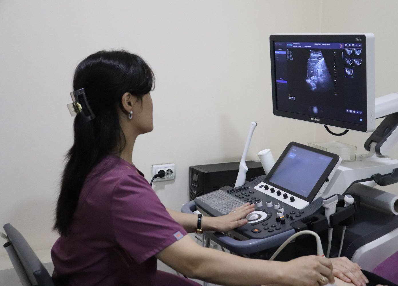
Our centre performs breast examinations on modern ultrasound equipment, and the result can be discussed with a gynecologist straight away so that next steps can be planned if needed. If you are unsure which method suits your age, talk it through with a doctor — it is a question that one visit settles.
Important: this article is for information only and does not replace a medical consultation. See a specialist for an accurate diagnosis and treatment plan.
Our doctors in this field