


All services
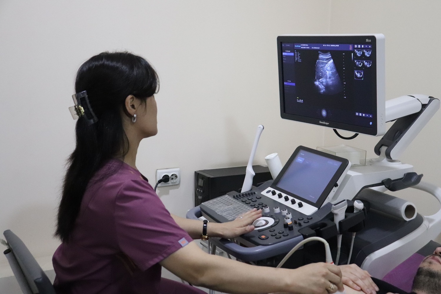
First trimester pregnancy ultrasound in Tashkent
A first trimester scan confirms that the pregnancy is in the womb, shows the heartbeat and gives an accurate due date, while at 11–13 weeks 6 days the first screening scan assesses ultrasound markers of chromosomal risk.
- Safe for the baby
- Report with measurements
- Partners welcome
- Duration20–40 minutes
- PreparationDepends on gestation
- SafetyNo radiation
- ResultsReport straight away
What the first trimester scan does
An early scan confirms that the pregnancy sac is inside the uterus and rules out an ectopic pregnancy — a dangerous condition that must be found early. From 6–7 weeks the heartbeat is visible, the number of babies is counted and, with twins, the type of placenta is identified. Dating by the baby’s length is the most accurate.
Between 11 weeks 0 days and 13 weeks 6 days the first screening scan is performed: nuchal translucency, the nasal bone, heart rate and early anatomy are assessed. Combined with blood markers, these measurements allow the risk of Down syndrome and other chromosomal conditions to be calculated. It is important not to miss this window.
- confirming a pregnancy in the uterus
- accurate dating
- seeing the fetal heartbeat
- detecting twins or more
- first screening at 11–13 weeks
Pregnancy scan schedule
Three routine screening scans are recommended during pregnancy, each with its own purpose:
- 11–13 weeks 6 days — first screening: crown–rump length, nuchal translucency and nasal bone, assessing the risk of chromosomal conditions
- 18–21 weeks — second screening: detailed anatomy of the baby’s organs and detection of structural anomalies
- 30–34 weeks — third screening: growth and position of the baby, placenta, amniotic fluid and blood flow
- extra scans may be ordered if the doctor considers them necessary
How the scan is done
Most scans are done through the abdomen: you lie on your back and gel is applied to the tummy. In very early pregnancy a transvaginal probe may be used for a clearer image. The doctor compares the measurements with norms for the stage of pregnancy and writes a report. Cutting down on gas-forming foods the day before is enough; fasting is not required.
Advantages of ultrasound
Ultrasound shows internal organs in real time and, unlike X-rays or CT, involves no ionising radiation. It can therefore be repeated as often as needed — including in pregnant women, babies and patients who require regular monitoring.
The scan is painless and needs no needles or injections. The probe glides over the skin with gel while the doctor assesses the size and structure of the organs and their blood flow on the screen. The report is issued immediately after the scan.
Ultrasound at Doctor Hasan Medical Center
Scans are performed by doctors trained in ultrasound diagnostics. The ultrasound room, laboratory and specialist doctors are in the same building, so you can show your result to a urologist, gynaecologist, endocrinologist or physician the same day and get a treatment plan straight away.
- the report and measurements are given right after the scan
- blood or urine tests can be done on site if needed
- booking in advance reduces waiting time
📖 Related article: Pregnancy Screening: The Schedule of Tests by Trimester
Frequently asked questions
Is it harmful to have frequent scans in pregnancy?
Is ultrasound harmful?
When will the result be ready?
Doctors who provide this service




Choose a convenient time or call us — our administrator will answer all your questions.
Other services in this area
- Second and third trimester pregnancy ultrasound
- Pregnancy ultrasound with fetal Doppler
- Fetal ultrasound with Doppler + 4D (colour photos, recording)
- 3D/4D fetal ultrasound
- All clinic services →
Important: the information on this page is for general guidance and does not replace an examination by a doctor. A specialist decides whether a test or treatment is needed.
📞 +998 95 146 00 72 · +998 95 146 00 72
✉️
🌐 Instagram · Telegram · Facebook · YouTube · TikTok