

All services
Kidney and adrenal gland ultrasound with Doppler in Tashkent
Measurement of blood flow in the renal arteries and veins is added to the kidney and adrenal scan. This helps find the cause of high blood pressure, especially in young patients or when medicines do not work.
- Painless, no radiation
- Detects stones and cysts
- Instant report
- Duration15–25 minutes
- PreparationLow-gas diet, full bladder
- PainNone
- ResultsImmediately after the scan
Why renal Doppler is needed
When a renal artery narrows, the kidney releases substances that raise blood pressure — this is renovascular hypertension. It often resists several medicines, and treating the artery can normalise pressure. Doppler measures flow velocity in the renal arteries and the resistive index of vessels inside the kidney to detect signs of narrowing.
The resistive index also reflects the state of kidney tissue in chronic kidney disease and diabetes. Renal vein thrombosis and the blood supply of kidney and adrenal masses are assessed too. The scan is done fasting after a low-gas diet and takes a little longer than a standard ultrasound.
- high blood pressure in young people
- hypertension not controlled by three or more medicines
- declining kidney function or rising creatinine
- assessing kidney vessels in diabetes
- vessels of kidney or adrenal masses
Preparation
The kidneys lie behind the abdominal cavity, close to the lower back. To see them clearly there should be little gas in the bowel and the bladder should be full:
- for 2–3 days cut down on gas-forming foods (beans, brown bread, cabbage, fizzy drinks)
- if the bladder is also scanned, drink 0.8–1 litre of still water an hour before and do not pass urine
- if the adrenal glands are examined, it is best not to eat for 6–8 hours
- bring previous ultrasound reports and urine test results
How the scan is done
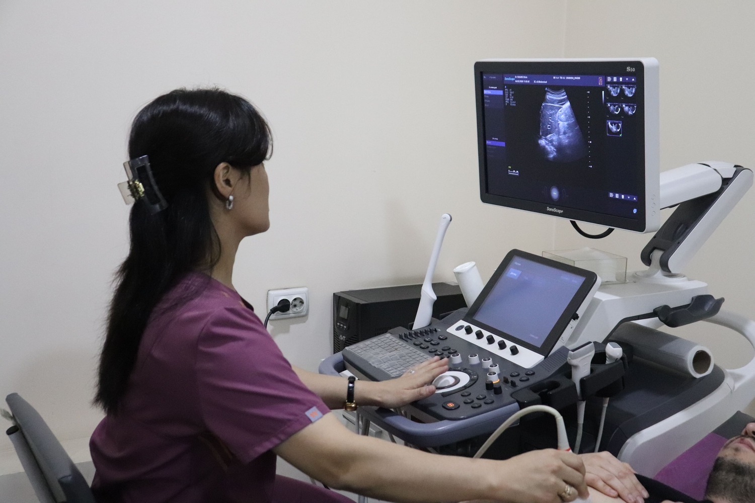
The kidneys are scanned from the back while you lie on your side or front, and sometimes from the front while lying on your back. The doctor assesses kidney size and position, cortical thickness, any widening of the calyces and pelvis, and stones, cysts or other lesions. After the full bladder has been viewed, you are often asked to pass urine so the residual volume can be measured.
Advantages of ultrasound
Ultrasound shows internal organs in real time and, unlike X-rays or CT, involves no ionising radiation. It can therefore be repeated as often as needed — including in pregnant women, babies and patients who require regular monitoring.
The scan is painless and needs no needles or injections. The probe glides over the skin with gel while the doctor assesses the size and structure of the organs and their blood flow on the screen. The report is issued immediately after the scan.
Ultrasound at Doctor Hasan Medical Center
Scans are performed by doctors trained in ultrasound diagnostics. The ultrasound room, laboratory and specialist doctors are in the same building, so you can show your result to a urologist, gynaecologist, endocrinologist or physician the same day and get a treatment plan straight away.
- the report and measurements are given right after the scan
- blood or urine tests can be done on site if needed
- booking in advance reduces waiting time
📖 Related article: Kidney Stones: Pain Attack, Ultrasound Diagnosis and Treatment
Frequently asked questions
Do I need to fast for a kidney ultrasound?
Is ultrasound harmful?
When will the result be ready?
Doctors who provide this service




Choose a convenient time or call us — our administrator will answer all your questions.
Other services in this area
- Kidney and bladder ultrasound
- Kidney and adrenal gland ultrasound
- Kidney, bladder and prostate ultrasound with Doppler
- Genitourinary ultrasound (kidneys, bladder, prostate)
- All clinic services →
Important: the information on this page is for general guidance and does not replace an examination by a doctor. A specialist decides whether a test or treatment is needed.
📞 +998 95 146 00 72 · +998 95 146 00 72
✉️
🌐 Instagram · Telegram · Facebook · YouTube · TikTok