

All services
Mediastinal lymph node ultrasound with Doppler in Tashkent
Doppler is added to the mediastinal node scan: the flow pattern in detected nodes is assessed, and they are reliably distinguished from the large vessels next to them.
- No preparation
- Assesses node structure
- Instant report
- Duration15–25 minutes
- PreparationFasting for deep nodes
- PainNone
- ResultsImmediately after the scan
Why Doppler helps in this area
Large arteries and veins run through the upper mediastinum, and on a standard scan a vessel seen in cross-section can look like a lymph node. Doppler shows the flow within a vessel, identifies the structure precisely and prevents mistakes.
In detected nodes, the flow pattern — through the hilum or chaotic — is an additional clue to their nature. Changes in flow during follow-up reflect the effect of treatment. For deep nodes, CT remains the main method.
- telling nodes apart from large vessels
- assessing the flow pattern in nodes
- documenting changes during follow-up
- detailed radiation-free assessment in children
- additional information alongside CT
How lymph nodes are assessed on ultrasound
Lymph nodes enlarge in response to infection and inflammation — a normal protective reaction. Besides size, the doctor assesses the node’s shape (oval or round), cortical thickness, whether the fatty hilum is preserved, and its internal structure. An oval shape with a preserved hilum usually indicates a reactive change, while a rounded node, loss of the hilum and uneven structure need further investigation.
Follow-up scans give more information than a single scan: nodes enlarged after an infection shrink within 4–6 weeks. If enlargement persists or increases, the doctor orders blood tests and other investigations.
- size and ratio of dimensions
- shape, outline and cortical thickness
- the hilum and internal structure
- relationship to surrounding tissues
- with Doppler — the pattern of blood flow
Advantages of ultrasound
Ultrasound shows internal organs in real time and, unlike X-rays or CT, involves no ionising radiation. It can therefore be repeated as often as needed — including in pregnant women, babies and patients who require regular monitoring.
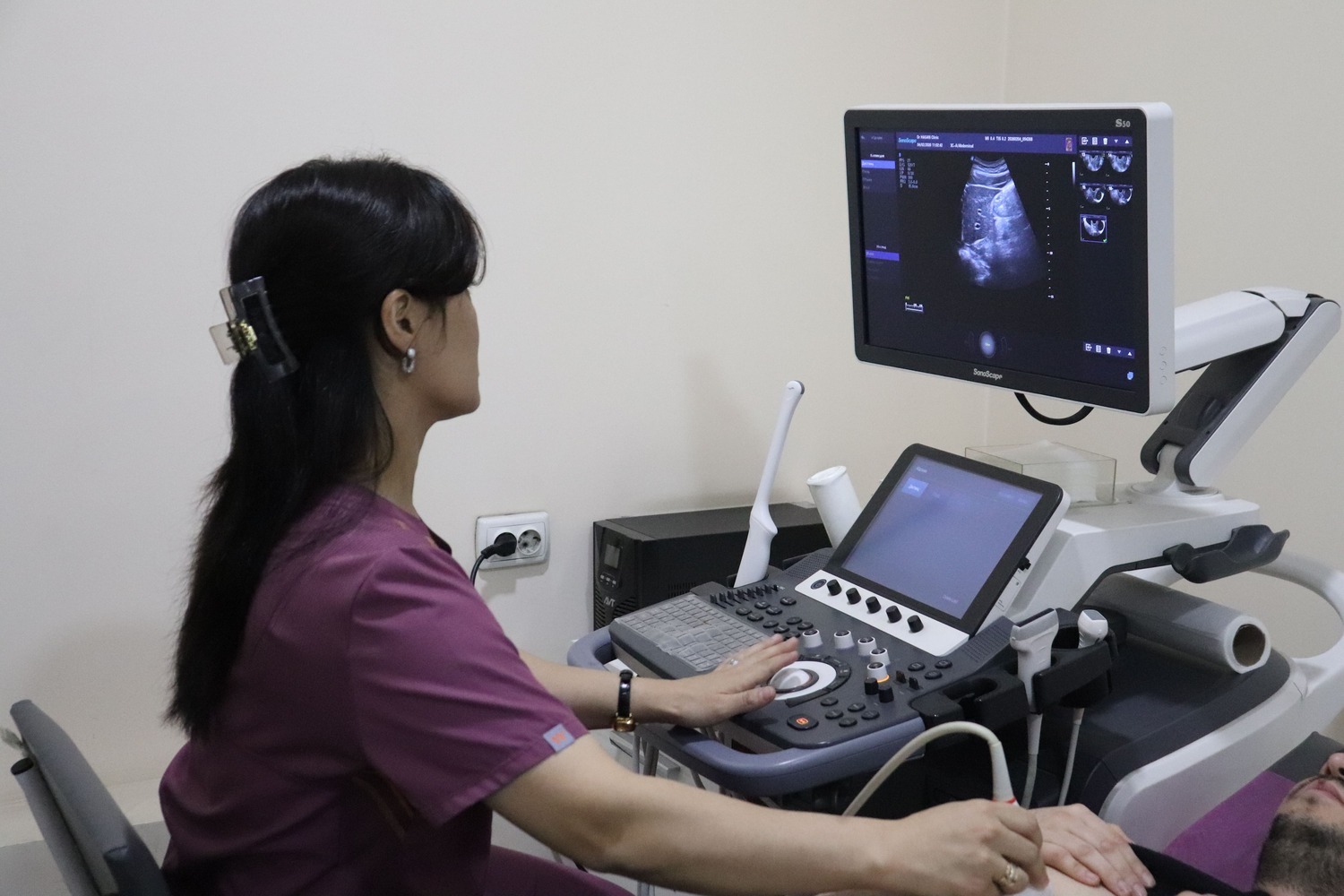
The scan is painless and needs no needles or injections. The probe glides over the skin with gel while the doctor assesses the size and structure of the organs and their blood flow on the screen. The report is issued immediately after the scan.
Ultrasound at Doctor Hasan Medical Center
Scans are performed by doctors trained in ultrasound diagnostics. The ultrasound room, laboratory and specialist doctors are in the same building, so you can show your result to a urologist, gynaecologist, endocrinologist or physician the same day and get a treatment plan straight away.
- the report and measurements are given right after the scan
- blood or urine tests can be done on site if needed
- booking in advance reduces waiting time
📖 Related article: Swollen Lymph Nodes: When It Is Normal and When to Investigate
Frequently asked questions
Is an enlarged lymph node always dangerous?
Is ultrasound harmful?
When will the result be ready?
Doctors who provide this service



Choose a convenient time or call us — our administrator will answer all your questions.
Other services in this area
- Lymph node ultrasound
- Lymph node ultrasound with Doppler
- Peripheral lymph node ultrasound
- Peripheral lymph node ultrasound with Doppler
- Mediastinal lymph node ultrasound
- Deep retroperitoneal lymph node ultrasound
- Retroperitoneal lymph node ultrasound with Doppler
- All clinic services →
Important: the information on this page is for general guidance and does not replace an examination by a doctor. A specialist decides whether a test or treatment is needed.
📞 +998 95 146 00 72 · +998 95 146 00 72
✉️
🌐 Instagram · Telegram · Facebook · YouTube · TikTok