

All services
Neurosonography with Doppler in Tashkent
Doppler is added to neurosonography: along with the baby’s brain structures, blood flow in the cerebral arteries and venous sinuses is measured. It gives extra information in oxygen shortage and suspected raised intracranial pressure.
- Through the fontanelle
- Safe for babies
- No preparation
- Duration10–15 minutes
- AgeWhile the fontanelle is open (usually up to 12–18 months)
- PainNone
- ResultsImmediately after the scan
What cerebral blood flow assessment adds
In babies born with oxygen shortage or prematurely, changes in brain blood flow can signal tissue damage in advance. Doppler measures flow velocity and the resistive index in the main cerebral arteries — values that reflect how well the brain is supplied with blood.
When intracranial pressure rises, arterial resistance increases, so the scan is useful in monitoring hydrocephalus. Venous flow is assessed to detect signs of venous congestion. The neurologist interprets the results together with the clinical examination.
- severe oxygen shortage and after resuscitation
- follow-up of premature babies
- suspected hydrocephalus or raised intracranial pressure
- tracking enlarged ventricles over time
- seizures and severe neurological signs
How neurosonography is performed
In babies the skull bones have not yet fully joined, and the soft spot between them — the fontanelle — acts as a natural window for ultrasound. The probe is placed gently on the fontanelle with gel and the doctor views the brain structures in different planes. The procedure is painless, involves no radiation and needs no anaesthesia.
Images are clearer when the baby is calm, so it is best timed after a feed or during sleep. Parents stay with the baby throughout. Once the anterior fontanelle closes, the scan becomes much less informative.
Preparing for the scan
No special preparation is needed, but these help keep your baby settled:
- feed the baby before the scan
- bring a nappy, a blanket and a favourite toy
- bring the maternity discharge summary and previous test results
- write down any concerns and questions from the neurologist
Advantages of ultrasound
Ultrasound shows internal organs in real time and, unlike X-rays or CT, involves no ionising radiation. It can therefore be repeated as often as needed — including in pregnant women, babies and patients who require regular monitoring.
The scan is painless and needs no needles or injections. The probe glides over the skin with gel while the doctor assesses the size and structure of the organs and their blood flow on the screen. The report is issued immediately after the scan.
Ultrasound at Doctor Hasan Medical Center
Scans are performed by doctors trained in ultrasound diagnostics. The ultrasound room, laboratory and specialist doctors are in the same building, so you can show your result to a urologist, gynaecologist, endocrinologist or physician the same day and get a treatment plan straight away.
- the report and measurements are given right after the scan
- blood or urine tests can be done on site if needed
- booking in advance reduces waiting time
Frequently asked questions
Up to what age can neurosonography be done?
Is ultrasound harmful?
When will the result be ready?
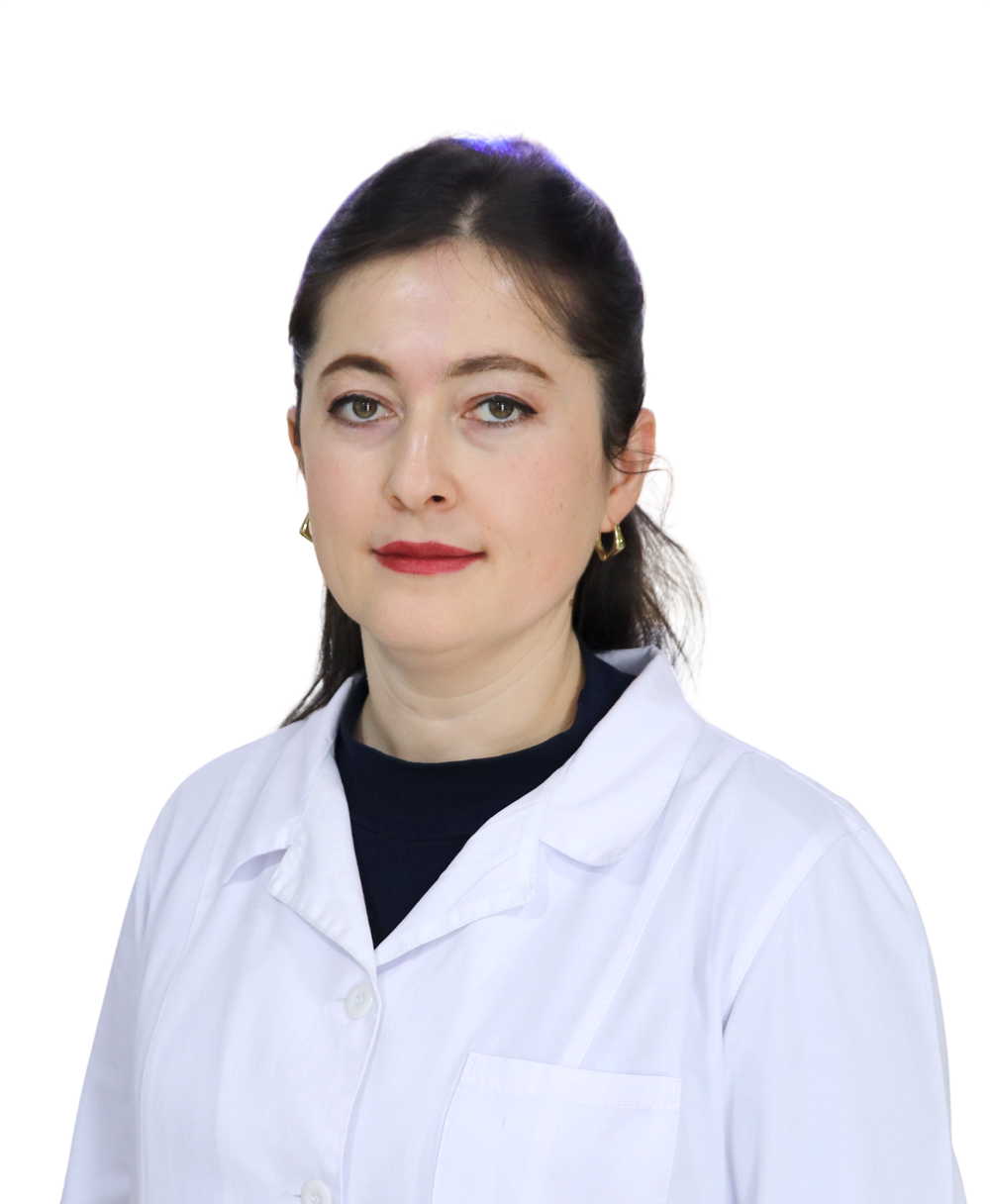
Doctors who provide this service



Choose a convenient time or call us — our administrator will answer all your questions.
Other services in this area
Important: the information on this page is for general guidance and does not replace an examination by a doctor. A specialist decides whether a test or treatment is needed.
📞 +998 95 146 00 72 · +998 95 146 00 72
✉️
🌐 Instagram · Telegram · Facebook · YouTube · TikTok