


Useful articles
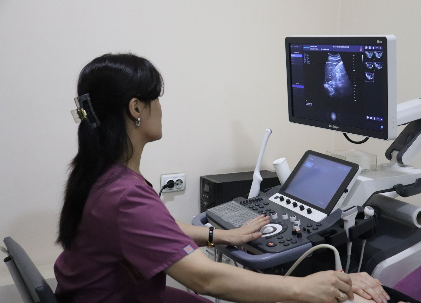
3D and 4D Ultrasound During Pregnancy: Dr.Hasan Medical Center
Ultrasound examination (US) during pregnancy is one of the most important methods for monitoring fetal development. Modern 3D and 4D ultrasound technologies provide a much clearer and more realistic view of the baby compared to standard ultrasound.
At Dr.Hasan Medical Center, advanced 3D and 4D ultrasound services are performed using modern equipment and supervised by experienced specialists 👨⚕️
What is 3D Ultrasound?
3D ultrasound creates a three-dimensional image of the fetus.
It allows doctors and parents to:
- clearly see facial features of the baby 👶
- observe the shape of hands and feet
- evaluate external fetal development in more detail
3D ultrasound gives parents a special opportunity to see their baby in a more realistic way for the first time.
🎥 What is 4D Ultrasound?
4D ultrasound is an advanced version of 3D imaging that adds real-time movement.
With 4D ultrasound, you can see:
- fetal movements 🤍
- hand and leg activity
- facial expressions (smiling, reactions, etc.)
It creates a live video-like experience of the baby inside the womb.
⏳ When is the Best Time to Do 3D and 4D Ultrasound?
The optimal period is:
- 24–32 weeks of pregnancy
During this time:
- the baby is well developed
- there is enough amniotic fluid for clear imaging
- fetal movements are more visible
🏥 3D and 4D Ultrasound at Dr.Hasan Medical Center
At Dr.Hasan Medical Center:
- modern ultrasound equipment is used
- experienced specialists perform the examination
- detailed fetal development assessment is provided
- 3D and 4D ultrasound services are available
This examination not only supports medical monitoring but also gives parents an emotional first “live meeting” with their baby 🤍
⚠️ Important Note
- 3D and 4D ultrasound are mainly advanced imaging methods
- standard ultrasound remains the primary diagnostic tool when medically necessary
- the procedure should be performed based on a doctor’s recommendation