

All services
Limb vein Doppler (venous duplex) in Tashkent
The state of the deep and superficial veins, how the vein valves work and whether there is a clot are assessed. It is the key test for varicose veins, leg swelling and heaviness, visible veins and suspected thrombosis.
- Colour flow mapping
- No preparation
- No radiation
- Duration20–40 minutes
- PreparationNone required
- PainNone
- ResultsImmediately after the scan
What a venous Doppler detects
Valves in the veins keep blood flowing only towards the heart. When they weaken, blood flows back down and the veins widen — leading to varicose veins and chronic venous insufficiency. Doppler shows which veins have faulty valves and where the backflow starts; this information is essential for choosing treatment and planning surgery.
Deep vein thrombosis is dangerous because a clot can break off and travel to the lungs. With sudden swelling and pain in a leg, the scan should not be delayed. The doctor presses on the vein with the probe to check that it collapses fully and assesses backflow with a straining test.
- enlarged veins and spider veins on the legs
- swelling and heaviness in the legs in the evening
- sudden swelling and pain in a leg (suspected clot)
- before varicose vein surgery or sclerotherapy
- bed-bound or post-operative patients
How Doppler works
When ultrasound bounces off moving blood cells its frequency changes (the Doppler effect). The machine converts this change into the direction and speed of blood flow. The vessel wall and any changes inside are seen in standard mode, and flow in colour and spectral modes — which is why the test is also called duplex (triplex) scanning.
The scan detects narrowing, plaques, clots, faulty vein valves and widened vessels. It needs no contrast and no radiation, so it can be repeated as often as necessary during follow-up.
Preparing for the scan
No special preparation is required, but the result is more accurate if you:
- avoid smoking and strong coffee for 2 hours beforehand — both affect vessel tone
- tell the doctor about any medicines that affect blood vessels
- remove jewellery if the neck vessels are being scanned
- bring previous Doppler reports
Advantages of ultrasound
Ultrasound shows internal organs in real time and, unlike X-rays or CT, involves no ionising radiation. It can therefore be repeated as often as needed — including in pregnant women, babies and patients who require regular monitoring.
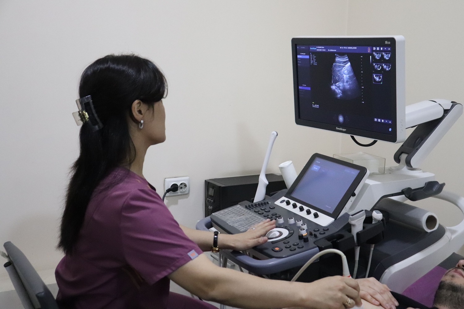
The scan is painless and needs no needles or injections. The probe glides over the skin with gel while the doctor assesses the size and structure of the organs and their blood flow on the screen. The report is issued immediately after the scan.
Ultrasound at Doctor Hasan Medical Center
Scans are performed by doctors trained in ultrasound diagnostics. The ultrasound room, laboratory and specialist doctors are in the same building, so you can show your result to a urologist, gynaecologist, endocrinologist or physician the same day and get a treatment plan straight away.
- the report and measurements are given right after the scan
- blood or urine tests can be done on site if needed
- booking in advance reduces waiting time
📖 Related article: Vascular Doppler Ultrasound: Who Needs It and When
Frequently asked questions
Is a Doppler scan painful?
Is ultrasound harmful?
When will the result be ready?
Doctors who provide this service



Choose a convenient time or call us — our administrator will answer all your questions.
Other services in this area
- Extracranial neck vessel Doppler
- Brachiocephalic artery Doppler (BCA)
- Upper limb arterial Doppler
- Lower limb arterial Doppler
- All clinic services →
Important: the information on this page is for general guidance and does not replace an examination by a doctor. A specialist decides whether a test or treatment is needed.
📞 +998 95 146 00 72 · +998 95 146 00 72
✉️
🌐 Instagram · Telegram · Facebook · YouTube · TikTok