


Useful articles
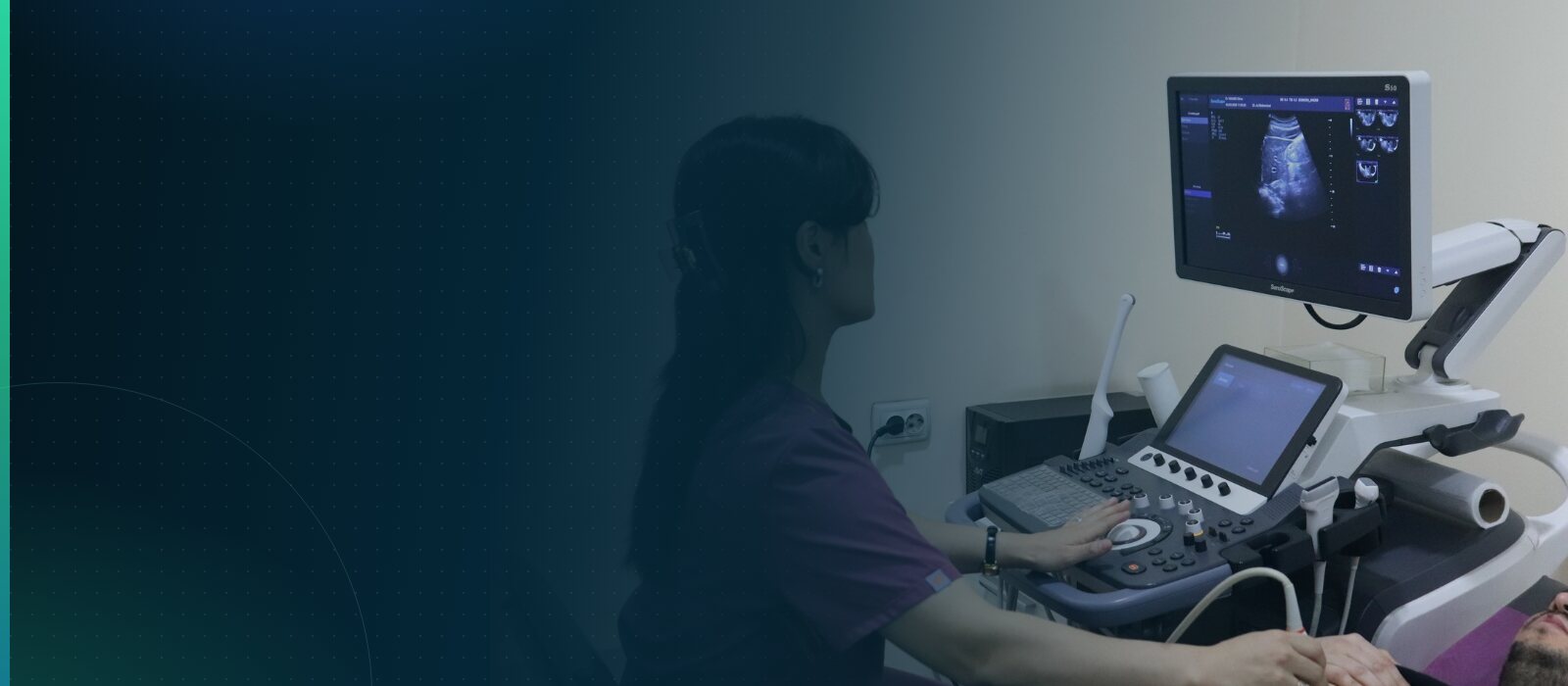
Kidney Ultrasound: Preparation, Normal Values and Results
A kidney ultrasound (renal ultrasound) is a painless, safe scan that shows the position, size and internal structure of the kidneys. It is usually the first test a doctor requests for loin pain, an abnormal urine test or high blood pressure. The report, however, often raises more questions than it answers: phrases such as “sand in the kidneys”, “dilated renal pelvis” or “thinned parenchyma” sound alarming, yet do not always mean disease. This article explains who needs a kidney ultrasound and when, how to prepare, what happens during the scan, the normal values for adults, what the most common wordings in a report mean, what ultrasound cannot show, and which symptoms should never wait for a planned scan.
- What a kidney ultrasound shows and who needs it
- How to prepare for a kidney ultrasound
- What happens during the scan and is it safe
- Normal values on a kidney ultrasound in adults
- Understanding the report: stones, “sand” and a dilated pelvis
- Limits of the method and how often to repeat it
- When to see a doctor urgently
- Frequently asked questions
- At Doctor Hasan Medical Center
What a kidney ultrasound shows and who needs it
Ultrasound shows the shape and size of the kidneys, the parenchyma (the working tissue) and the collecting system, as well as stones, cysts and other masses. The bladder is usually examined at the same time. A doctor requests the scan for:
- pain in the loin or side;
- blood, protein or white cells found on urinalysis;
- high blood pressure, especially in a young person or when it is hard to control;
- swelling, or a raised blood creatinine;
- recurrent urinary tract infections;
- diabetes — to keep an eye on the kidneys;
- follow-up of a known stone or cyst, or an injury to the back;
- in children — fever with no clear cause, or bedwetting.
How to prepare for a kidney ultrasound
If only the kidneys are being examined, no special preparation is needed and you may eat as usual. If you are prone to marked bloating, avoiding gas-forming foods (pulses, cabbage, dark bread, fizzy drinks) for a day beforehand helps, because bowel gas blurs the image.
If the bladder (and, in men, the prostate) is examined too, come with a full bladder: drink 0.5–1 litre of still water about an hour before the scan and do not pass urine. Bring earlier scan reports and test results — comparing them over time makes the diagnosis easier.
What happens during the scan and is it safe
You lie first on your back and then on your side; the doctor spreads gel on the skin and moves the probe over the loin and abdomen. You will be asked to take a deep breath and hold it briefly so that the kidneys can be seen more clearly. The scan takes 10–20 minutes and is painless. There is no radiation, so it is safe in pregnancy and childhood and can be repeated whenever necessary. If the bladder has been examined, you empty it and the doctor then measures the residual urine. When a dropped kidney (nephroptosis) is suspected, the kidney is also checked with you standing.
Normal values on a kidney ultrasound in adults
The figures below are a guide for adults; they vary a little with height, age and build.
| Measurement | Normal |
|---|---|
| Length | 10–12 cm |
| Width and thickness | 5–6 cm and 4–5 cm |
| Parenchymal thickness | 15–25 mm, thinner with age |
| Difference in size between the two kidneys | Under 1.5–2 cm |
| Residual urine | Under 50 ml |
In addition, a healthy kidney has a smooth outline, a renal pelvis that is not dilated, and no stones.
Understanding the report: stones, “sand” and a dilated pelvis
An ultrasound report describes what was seen; it is not a finished diagnosis. The most common wordings are:
| Wording in the report | What it means | Next step |
|---|---|---|
| Stone (calculus): a bright focus with a shadow | Kidney stone disease; tiny stones and stones in the ureter can be missed | Urologist; CT is the reference test if confirmation is needed |
| “Sand”, microliths, salt diathesis | A vague wording, not a diagnosis | Confirm with a urine test |
| Dilated pelvis, hydronephrosis | An obstruction to urine flow; mild right-sided dilation is common in pregnancy | Find the cause; urology review |
| Cyst | A simple cyst is harmless; a complex one needs clarifying | Monitoring, or CT/MRI |
| Solid mass | A tumour has to be ruled out | Contrast CT or MRI |
A note about “sand” or “salt diathesis” does not yet mean kidney stones, and a simple kidney cyst usually needs nothing more than monitoring. In pyelonephritis the scan is often normal — the diagnosis rests on symptoms and urine tests. Small kidneys with thin, bright parenchyma are typical of chronic kidney disease. A horseshoe kidney, a duplex collecting system or a single kidney are anatomical variants that more often need to be noted than treated.
Limits of the method and how often to repeat it
Ultrasound shows the structure of the kidneys, not their function. To know how well the kidneys are filtering the blood, blood creatinine (from which the filtration rate, eGFR, is estimated) and a urine test are needed: the picture can look normal even when function is reduced. Excess weight and bowel gas lower image quality, and tiny stones may go unnoticed.
Doppler of the renal vessels assesses the blood supply; it is requested when narrowing of a renal artery is suspected in someone with high blood pressure.
The scan is repeated when there is a reason: stones are usually checked every 6–12 months, cysts according to their category. A healthy adult with no symptoms or risk factors does not need a yearly kidney ultrasound “just in case”.
When to see a doctor urgently
In the following situations do not wait for a planned scan — see a doctor or call an ambulance the same day:
- severe pain in the loin or side that comes in waves (renal colic);
- loin pain with fever and chills;
- visible blood in the urine;
- very little urine or none at all;
- pain after an injury to the back or abdomen.
Frequently asked questions
Can I eat and drink before a kidney ultrasound?
Yes. If only the kidneys are being scanned, you do not need to fast. If you tend to bloat badly, it helps to avoid gas-forming foods for a day beforehand. If the bladder or prostate will be examined as well, drink 0.5–1 litre of still water about an hour before the scan and do not pass urine: with a full bladder the doctor can assess its wall and the residual urine more accurately.
What does “sand in the kidneys” mean on a report?
“Sand”, “microliths” and “salt diathesis” are not diagnoses but vague descriptions of tiny bright dots on the image, which ultrasound cannot reliably tell apart. The finding should be checked with a urine test, which looks for salt crystals, blood and white cells. If you have no symptoms and the tests are normal, drinking enough water and routine follow-up are usually sufficient; do not take “sand-flushing” remedies on your own.
If my kidney ultrasound is normal, are my kidneys healthy?
Not necessarily. Ultrasound shows the structure of the kidneys, not how well they work: in early chronic kidney disease the picture can be normal, and in pyelonephritis the scan is often unchanged too. Function is assessed by blood creatinine and a urine test. That is why people with diabetes or high blood pressure are advised to have these tests alongside the scan.
Is a dilated renal pelvis in pregnancy dangerous?
Mild dilation of the renal pelvis, especially on the right, is common in pregnancy: the growing womb presses on the ureter and hormones relax its wall. If there is no pain, no fever and the urine tests are normal, monitoring is usually all that is needed, and it settles after the birth. If you develop loin pain, a temperature or burning when passing urine, see a doctor without delay.
At Doctor Hasan Medical Center
The centre offers a kidney and bladder ultrasound, a kidney and adrenal ultrasound with Doppler and a combined genitourinary scan (kidneys, bladder and prostate). A urinalysis can be done here as well when needed. The ultrasound doctor describes in detail the size of the kidneys, the parenchyma and renal pelvis, and any stones or cysts found. The result can be discussed the same day at a urologist consultation: the doctor sets the report against your symptoms and test results, explains it in plain language and draws up a personal follow-up schedule. If hospital treatment or surgery is needed, the doctor will discuss the options and next steps. Please remember to bring your earlier reports.
Important: this article is for information only and does not replace a medical consultation. See a specialist for an accurate diagnosis and treatment plan.
Our doctors in this field